Deep learning can help to detect life-threatening cerebral aneurysms on CT angiography
Deep learning (DL) can help physicians detect potentially life-threatening cerebral aneurysms on CT angiography. Researchers from Stanford University have developed an artificial intelligence (AI) tool that can help radiologists improve their diagnoses of brain aneurysms by highlighting the areas of a brain scan that are likely to contain an aneurysm.
Cerebral aneurysms are weakened areas of blood vessels in the brain. Deep learning offers tremendous potential as a supplementary tool for a more accurate interpretation of cerebral aneurysms.
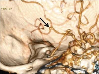
Cerebral aneurysms are weakened areas of blood vessels in the brain. Left untreated, they can leak or rupture, with sometimes fatal results. To overcome the inherent challenges of complex neurovascular anatomy and the potentially fatal outcome of a missed aneurysm, the researchers focused on creating an AI tool that could accurately process large stacks of 3D images and complement clinical diagnostic practice. Detection and characterization of these aneurysms are critical, as the risk of rupture depends on the size, shape, and location of the aneurysm. The researchers focused on its ability to identify the presence of aneurysms rather than on detecting their absence.
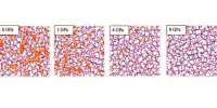
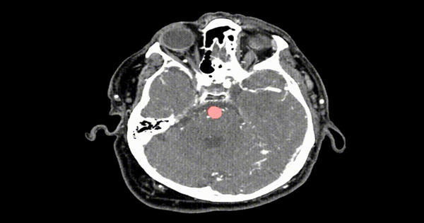
Following the training, the algorithm decides for each voxel of a scan whether there is an aneurysm present. CT angiography is usually the first choice for evaluating cerebral aneurysms. As a result of the HeadXNet tool, the algorithm’s conclusions overlaid as a semi-transparent highlight on top of the scan. The exam is highly accurate, but cerebral aneurysms can be overlooked on the initial assessment due to their small size and the complexity of the blood vessels in the brain.
“In our daily work we are always faced with cases in which some important lessons have been missed by the human eye,” said study senior author Xi Long, Ph.D., from the Department of Radiology at Tongji Medical College’s Union Hospital in Wuhan, China. “Cerebral aneurysms are among those small lesions that may be overlooked on the routine assessment of radiological images.”
Deep learning offers tremendous potential as a supplementary tool for a more accurate interpretation of cerebral aneurysms. A deep learning system is trained on existing images and learns to recognize abnormalities that can be difficult for a human observer to see. In radiology, deep learning has been recently been used in a variety of roles assisting radiologists, such as in the detection of tuberculosis on chest X-rays.
AI Helps Detect Brain Aneurysms on CT Scans
In the new study, Dr. Long and colleagues developed a fully automated, highly sensitive algorithm for the detection of cerebral aneurysms on CT angiography images. They used CT angiograms from more than 500 patients to train the deep learning system, and then they tested it on another 534 CT angiograms that included 649 aneurysms.
The algorithm detected 633 of the 649 cerebral aneurysms for a sensitivity of 97.5%. It also found eight new aneurysms that were overlooked in the initial assessment. Statistical analysis revealed that deep learning assistance enhanced radiologists’ performance. The improvement was most pronounced in the less experienced radiologists.
Cerebral aneurysms are weakened areas of blood vessels in the brain. If left untreated, they can leak or burst, with sometimes fatal consequences. Detection and characterization of these aneurysms are critical because the risk of rupture depends on the size, shape, and location of the aneurysm.
“The developed deep-learning system has shown excellent performance in detecting aneurysms,” Dr. Long said. “We found some aneurysms that were overlooked by the human readers on the initial reports, but they were successfully depicted by the deep-learning system.”
The system has some limitations, Dr. Long noted. It can miss very small aneurysms or aneurysms located close to similar density structures like bones. It also suffers from false-positive results, meaning that it mistakenly identifies structures similar to aneurysms as aneurysms, which necessitates careful revision of the system suggestions by human readers.
“Simply put, the deep-learning system is intended to assist human readers, not to replace them,” Dr. Long said.
The system needs further validation on more heterogeneous data, such as images from people in different parts of the world, which is essential for assessing its generalizability and applicability to daily clinical work.
The statistical analysis showed that the support of deep learning improved the radiologist’s performance. The improvement was most pronounced among the less experienced radiologists.