Nucleocapsid as a novel drug for targeting the shell of Ebola virus
Ebola is a highly virulent pathogen causing severe hemorrhagic fever with a high case fatality rate in humans and non-human primates (NHPs). As the world grapples with the coronavirus (COVID-19) pandemic, another virus has been raging again in recent months: Ebola. Researchers are using supercomputers to simulate the inner workings of Ebola (as well as COVID-19), looking at how molecules move, atom by atom, to carry out their functions. Since the first terrifying outbreak in 2013, the Ebola virus has periodically emerged in Africa, causing horrific bleeding in its victims and, in many cases, death.
Research teams led by Professor Juan Perilla at the University of Delaware are using supercomputers to simulate the inner workings of Ebola, observing the way molecules move, atom by atom, to carry out their functions. Perilla and his team of graduate and undergraduate students in UD’s Department of Chemistry and Biochemistry are using supercomputers to simulate the inner workings of Ebola, observing the way molecules move, atom by atom, to carry out their functions. In the team’s latest work, they reveal structural features of the virus’s coiled protein shell or nucleocapsid, which may be promising therapeutic targets, more easily destabilized and knocked out by antiviral treatment. Now, they have revealed structural features of the Ebola virus’s protein shell to provide therapeutic targets to destabilize the virus and knock it out with antiviral treatment.
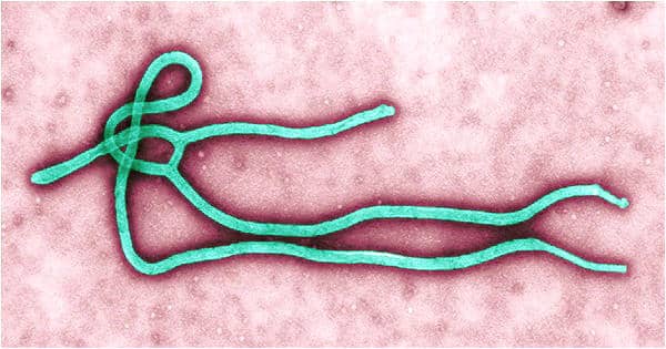
“The Ebola nucleocapsid looks like a Slinky walking spring, whose neighboring rings are connected,” Perilla said. “We tried to find what factors control the stability of this spring in our computer simulations.”
The life cycle of Ebola is highly dependent on this coiled nucleocapsid, which surrounds the virus’s genetic material consisting of a single strand of ribonucleic acid (ssRNA). Using supercomputer simulations researchers reveal that the structural stability of the Ebola nucleocapsid is dependent on the presence of RNA and interactions with charged ions. Nucleoproteins protect this RNA from being recognized by cellular defense mechanisms. Through interactions with different viral proteins, such as VP24 and VP30, these nucleoproteins form a minimal functional unit — a copy machine — for viral transcription and replication.
While nucleoproteins are important to the nucleocapsid’s stability, the team’s most surprising finding, Perilla said, is that in the absence of single-stranded RNA, the nucleocapsid quickly becomes disordered. But RNA alone is not sufficient to stabilize it. The team also observed charged ions binding to the nucleocapsid, which may reveal where other important cellular factors bind and stabilize the structure during the virus’s life cycle.
Perilla compared the team’s work to a search for molecular “knobs” that control the nucleocapsid’s stability like volume control knobs that can be turned up to hinder virus replication.
The UD team built two molecular dynamics systems of the Ebola nucleocapsid for their study. One included single-stranded RNA; the other contained only the nucleoprotein. The systems were then simulated using the Texas Advanced Computing Center’s Frontera supercomputer — the largest academic supercomputer in the world. The simulations took about two months to complete.
Perilla commented that their new understanding of the nucleocapsid, the most abundant protein in the virus, and a highly immunogenic component, “may facilitate the development of new antiviral treatments”.
Ebola virus infection in humans generally occurs through direct contact with mucosal surfaces, skin abrasions, or contaminated needles. Currently, Perilla and Jodi Hadden-Perilla are using supercomputer simulations to study the novel coronavirus that causes COVID-19. Although the structures of the nucleocapsid in Ebola and COVID-19 share some similarities — both are rod-like helical protofilaments and both are involved in the replication, transcription, and packing of viral genomes — that is where the similarities end.


