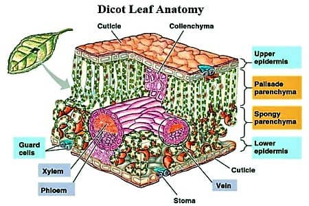
Dicot is a term used to explain a group of flowering plants that have two seed leaves. It generally has secondary growth that shows up like wood and bark in their stems.
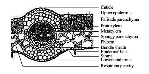
The anatomical structure of a dicot leaf / dorsiventral leaf. The following arrangement of tissues are seen in the cross-section of a dorsiventral leaf
- Upper epidermis
- Mesophyll tissue
- Vascular tissue
- Lower epidermis.
(1) Upper epidermis: It is the outermost layer and secretes a waxy substance called the cuticle. This is a single layer of cells containing few or no chloroplasts. In floating aquatic plants such as Nymphaea, the stomata are present on the upper epidermis. The cuticle helps retain water inside the leaf cells.
- It is composed of one layer of compactly arranged parenchymatous cells.
- A thick cuticle layer is present on the upper epidermis. The outer wall of the cells are thick and covered with a thick layer of cuticle.
- Epidermal cells are devoid of chloroplasts. Stomata are generally absent in the upper epidermis.
- Chlorophyll and stomata are absent in this layer.
Functions: (i) It protects the inner tissues, (ii) Saves the inner use of water.
(2) Mesophyll tissue: Ail the tissues in between upper and lower epidermis except for veins and the branches of veins are called mesophyll tissue. It is the chloroplast containing a portion of the leaf.
It has two types of tissues:
(a) Palisade parenchyma
(b) Spongy parenchyma
Palisade parenchyma: Below upper epidermis 2-3 rows of long parenchymatous cells are present which is enriched with plenty of chlorophyll. They are mainly responsible for producing food and oxygen for the plant through photosynthesis.
- Palisade tissue is present on the upper (dorsal or adaxial) surface of the leaf.
- Composed of usually one or two layers of cells, and cells are compactly packed without any intercellular spaces.
- Contain a large number of chloroplasts.
Function: To prepare food, the main function of palisade tissue is to perform the photosynthesis.
Spongy parenchyma: Spongy tissue occupies below the palisade tissue and cells are loosely arranged and irregularly shaped. Cells of spongy tissue are in contact with the atmosphere through the stomata. The cells in this layer contain few chloroplasts and are therefore not generally responsible for photosynthesis.
- The round or oval-shaped cells, situated below the palisade parenchyma, which are arranged scattered are said to be spongy parenchyma
- Within the cells, great deals of inter-cellular spaces are present.
- A little chloroplast is present m these cells
- Cells of the spongy tissue contain chloroplasts; however, the amount of chloroplasts is less than that of palisade tissue.
Functions: The main function of spongy tissue is to perform a gaseous exchange. (Absorption of Carbon Dioxide (CO2) and release of Oxygen (O2) and water vapor) and prepare food.
(3) Vascular bundle: Vascular tissue in the leaves is called vein. The pattern of the vein arrangement is called venation. Dicots have reticulate (net-like) venation.
- Veins and their branches make the vascular bundles.
- Vascular bundles consist of xylem and phloem and they are the conjoint, collateral and closed type.
- Vascular bundles are surrounded by one layer of parenchymatous cells called bundle sheath.
- Vascular tissue is irregularly distributed in the mesophyll.
- Each vascular bundle is surrounded by parenchymatous bundle sheath or border parenchyma.
Functions: To conduct water and prepared food materials.
(4) Lower epidermis: The lower epidermis is similar to the upper epidermis. They usually composed of a single layer of compactly packed parenchymatous cells. They are responsible for the transport of water and food.
- It is composed of one layer of compactly arranged parenchymatous cells.
- It contains a thin cuticle layer around this laser.
- Cuticle present, but relatively thinner than that of the upper epidermis.
- Numerous stomata are present on the lower epidermis.
Functions: (i) To protect the inner tissues (ii) Takes part in transpiration and gaseous exchange.