Magnetic Resonance Imaging (MRI) is a process of determining diseases by magnetic resonance, which is performed in 15 to 90 minute according to part of the body.
Principle
In an MRI machine, when current is started flowing in the wire, a strong magnetic field is created. Another coil of the magnet then sends or receives the radio wave. Consequently, protons of the body become excited by absorbing radio wave. Excited molecule sends the signal to the computer of the MRI machine by releasing excess energy, which captures the image of the related part of the body in the movable carbine. From the image, the disease can be determined.
Process
Using the property of magnet in atom, magnetic resonance of signal of the nucleus is captured 70% water exists in our body cell. The hydrogen atom (1H1) of water is an NMR active atom. ‘H’ atom works as a small magnet and spins in its own axis.
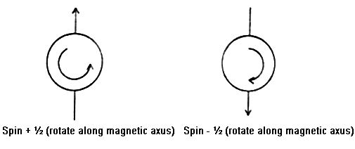
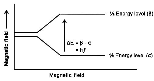
If the compound of hydrogen atom sets in a strong magnetic field, H nucleus splits into two energy level. Value of energy level which is along magnetic field is lower and level which is opposite to magnetic field is called higher energy state (β—level) For general reason more H —nucleus is situated in α—level. In this test, when electrical magnetic radiation is sent in the sample, H—nucleus in the lower level absorbs energy and transferred to the higher level (β—level) and give a signal by establishing resonance.